Jelenlegi hely
Transforming CT studies of the pelvic region to a common reference frame
GE Medical System
Our automatic registration method was tailored to the needs of a model-based segmentation framework of the pelvic area in CT studies in collaboration with GE Medical Systems. Registration was used as a preprocesing task. The goal of it was the acceptable alignment of the pubic bone area (where the two organs of interest, the prostate and bladder are located) of studies from different patients.
The method was used in the following two tasks.
- The collected CT studies were transformed to a common reference frame by selecting a suitable study and using it as a reference. Then a statistical atlas was computed which shows how probable it is that the given voxel belongs to a specific organ for each voxel of the reference frame. Manual segmentation was necessary to build the atlas, which was carried out by three experts.
- The second task occurs in the clinical software, where the statistical atlas information is transformed to the coordinate system of the image to be segmented for initialization of the segmentation method. A deformable organ model could also be used instead of the statistical atlas. The scope of our discussion was the role of registration as a preprocessing step, we did not deal with deformable organ models at this point.
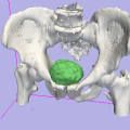
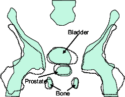
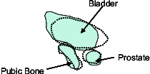
The applied algorithm consists of two steps. The idea is that after a transformation having translational, rotational and non-uniform scaling parameters which gives global optimal alignment, a refinement step is performed. Global registration prefers alignment of body parts of big volume, like spine or pelvis, causing slight or big differences in the pubic bone area. It is assumed that scale parameters are satisfactorily determined by this global part (Figure 1). During refinement, scale parameters are kept from the global part, then an optimal rigid-body transformation is searched in the pubic bone region only (Figure 2). The refinement requires the manual selection of the local neighborhood of the pubic bone in the reference study.


It was shown that our proposed two-step method (local refinement in the pubic bone area after a global registration) takes prostate regions significantly closer to each other when transforming to the common reference frame. GE Medical Systems provided us with an image database of 26 CT studies and their expert segmented prostate and bladder regions. In our database the failure rate of registrations was low (three out of 26) even though the studies were "real-life", many of them distorted by metallic objects, or not satisfying the assumed protocol (e.g., wrong patient position, contrast agent is visible in the images). Utilizing several optimizations (e.g., by using only the coarser levels of a hiearchical representation), the running time is currently between 20-40 seconds on a 3 GHz Pentium IV desktop PC. It can be further reduced to nearly its half by reducing the number of voxels in the reference volume removing the unnecessary parts (e.g., using the bounding box of the patient data). Figure 3 shows a result after automatic initial organ placement.
